Some eye conditions which concern the front of the eye can be treated simply, using eye drops, lid hygiene and tear nutrient therapy. But there are many eye problems which need more.
The eye is made up of several structures, that come together to bring vision. The front windscreen of your eye is called the cornea- it is a clear structure without any blood vessels, through which you see the iris (the coloured part) and the pupil (a hole in the iris- seen as black). Around the cornea, a clear film covers the white of the eye- this is called the conjunctiva. The conjunctiva covers all areas from the cornea to the inside of the eyelids.
If the cornea or conjunctiva is damaged it can cause pain and reduced vision (blur). When this happens it is very important to restore these structures to their normal state as quickly as possible.
Eye problems which may be requiring treatment include keratitis, corneal scars, chemical burns, corneal defects, recurrent erosion syndrome and severe dry eye syndrome.
One treatment option which may make a huge difference to these patients is the use of amniotic membranes.
This short summary will explore what it is, how it works, research into prevention and treatment and safety of amniotic membranes.
What is an amniotic membrane?
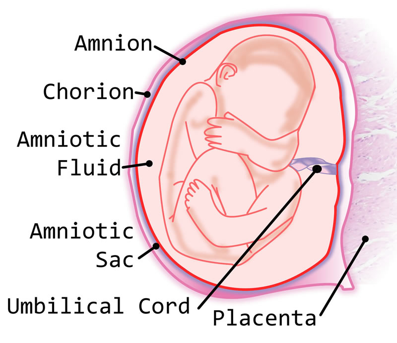
Amniotic membranes used in optometry come from the placenta of a women during a prearranged caesarean birth.
The amniotic membrane exhibits therapeutic actions to help a baby to develop in the womb. These healing properties can be utilised to help the eye surface to repair itself.
How does an amniotic membrane work?
Amniotic membranes work to reduce inflammation and promote quicker healing, meaning less pain and scarring to the patient.
What does the research say?
Amniotic membranes have been used in the USA for over 20 years. Research shows amniotic membranes have antimicrobial properties. Amniotic membranes can be implanted without risk of rejection as it does not contain cell-fighting cells.
Is it safe? Is it human amniotic membrane?
The membrane is harvested during elective caesearian section in a sterile environment. Donors are screened for all transmissible diseases before harvesting and the graft amniotic membrane tissue is tested again before being processed. Also the amniotic membrane is treated with broad spectrum antibiotics after collection.
There are two types of amniotic membrane used in optometry. Cryopreserved (frozen) membranes are kept at a low temperature and may be secured by a ring or band. The band maybe uncomfortable The dehydrated type is preserved using a vacuum and is limited to wound coverage.
How is the procedure carried out?
Dehydrated amniotic membranes are packaged dry. Anaesthetic drops are placed in the eye as an eye drop. The membrane is placed on the cornea ensuring the structures are the correct way round. Saline drops are added and a 'bandage' contact lens is added to cover the membrane and the corneal surface. After 7 days the membrane has dissolved and the contact lens is removed.