Matheson Optometrists now has invested the Zeiss Cirrus HD-OCT, Optical Coherence Tomography (OCT) available in all of our practices. It Utilises Spectral Domain (also known as Fourier Domain or transform OCT), Optical Coherence Tomography (OCT) rather than the earlier more common Time Domain technology. Spectral Domain is 60-70 times faster and so is less affected by such things as eye movement artifacts. It uses a broader band light source with improved depth resolution and aquisition speed. Andrew Matheson believes in using the best, most up-to-date technology to help detect eye conditions as early as possible. To make this happen we try and keep our charges for using this technology as low as reasonably possible. This is reflected in our private charge for providing an OCT scan of only £48.00. (See our fees)
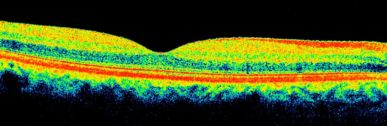
The Zeiss Cirrus HD-OCT, is a non-invasive technology originallyused for imaging the retina - the multi-layered sensory tissue lining the back of the eye. It is particularly good at picking up the early signs of Age Related Macula Degeneration in the early stages when treatment is most successful.
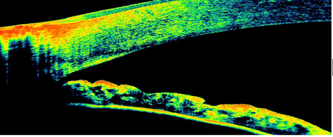
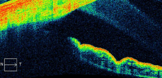
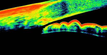
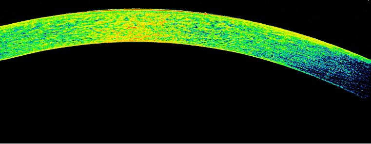
The Optical Coherence Tomography (OCT) scanner also is used to visualise the anterior chamber angle structures that can be affected in Narrow Angle Glaucoma, Plateau Iris Glaucoma and Pigment Dispersion Glaucoma, among other conditions.
Normal Anterior ChamberDrainage Angle
Wide Open Anterior Chamber Drainage Angle
Narrow Anterior Chamber Drainage Angle
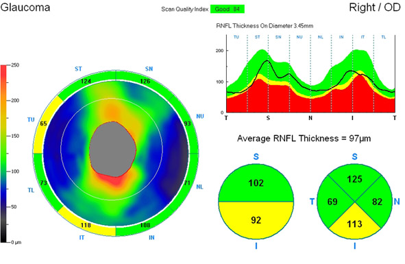
The Optical Coherence Tomography (OCT) scanner is further used in assessing the thickness of the Retinal Nerve Fibre Layer adjacent to the Optic Disc (Optic Nerve Head). This gives us a measurement and a map of Retinal Nerve Fibre Layer thickness. This will become our base for accurate quantitative data for more efficient long term monitoring for early signs of Glaucoma. In Glaucoma and some other conditions, the nerve fibres leaving the eye via the optic nerve head die off and thus assessing the thickness of this nerve fibre layer can often show the early signs of Glaucoma before the damage can be demonstrated by more convential methods such as Visual field Testing.
Also used in Glaucoma assessment we utilise the Optical Coherence Tomography (OCT) scanner to measure the Centre Corneal Thickness, which gives us a measurement which relates to corneal rigidity or stiffness. Central corneal thickness can affect the readings optometrists and ophthalmologists obtain by tonometry (intra-ocular pressure (IOP) measurement), because a thick and stiffer cornea will give an artificially elevated intra-ocular pressure (IOP) reading. Conversely, a thin and less rigid cornea will produce a reading that is actually lower than the true pressure. Glaucoma may thus go undetected in people with thin a cornea or might be misdiagnosed in people with a thicker cornea. In addition, having a thin cornea is an independant risk factor for Glaucoma as it may mean that the Optic Nerve Head is more vunerable to Glaucoma damage
Glaucoma may thus go undetected in people with thin a cornea or might be mis-diagnosed in people with a thicker cornea. In addition, having a thin cornea is an independant risk factor for Glaucoma as it may mean that the Optic Nerve Head is more vunerable to Glaucoma damage.
With its advanced, next generation, precise algorithms, The Zeiss Cirrus provides Matheson Optometrists with highly detailed, high-definition maps and images of the macula and retinal structures. This allows us to make the most accurate diagnosis possible.
One of the other important benefits of this form of retinal scanning is that being able to scan the various layers of the retina not normally visible when an optometrist or ophthalmologist can let us detect the early signs of conditions such as Wet Macula Degeneration, Epiretinal Membrane and Macular Holes, often before it is visible by conventional methods. Because the more common Dry Macular Degeneration can change to the more agressive and nowadays, often treatable Wet Macular Degeneration, screening patients with dry macular degeneration with Optical Coherence Tomagraphy (OCT) can often pick up this change in type early enough for treatment to be started effectively.