Vitreomacular traction or VMT for short, is a condition in which shrinkage of the vitreous jelly within the eye pulls on the central macular area of retina. This causes central visual impairment, either in the form of a generalised blurring or distortion.
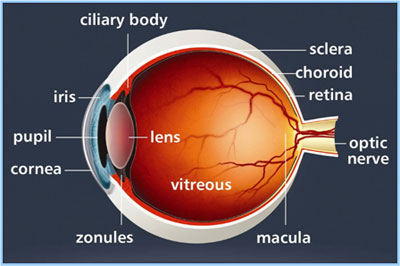
When light enters the eye it passes through the various tissues to reach the retina. The pathway of light starts at the tear film passing through the cornea, aqueous humour before the iris (the coloured part of the eye), crystalline lens, vitreous humour and finally the inner layers of the retina. The vitreous humour is a jelly-like substance which fills the majority of the posterior space in the eyeball. This jelly is attached to the retina at the optic nerve, macula and periphery called the ora serata.
As we age the jelly-like vitreous contracts and and degenerates, and becomes more liquid. This can lead to ‘floaters’ appearing in the patients vision. As this progresses the vitreous jelly often separates from the retina. when it does so completely it is called a Posterior Vitreous Detachment. In rare cases this when it does not fully detach, the jelly can tug on parts the retina, causing traction damage.
When the jelly pulls on the reina where it is thinnest at the Ora serrata, this can lead to holes and tears forming in the peripheral retina leading to a Retinal Detachment.
Sometimes the vitreous jelly fails to release from the macular region. Vitreomacular traction is the term used to describe the vitreous as it pulls on the central macula area. This can result in distortion and/or blurring of vision.
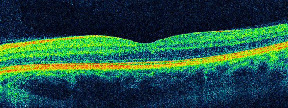
OCT scans can image the layers of the retina so that we are able to visualise the effect vitreomacular traction has on the structures.
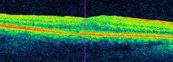
In a typical unaffected patient the layers of the retina have the regular appearance as below:-
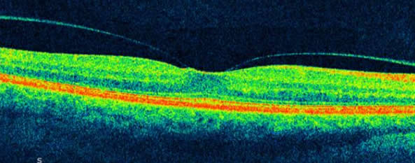
In an eye affected by early stages of partial vitreous separation you can see Vitreo Macular Adhesion and slight traction starting:-
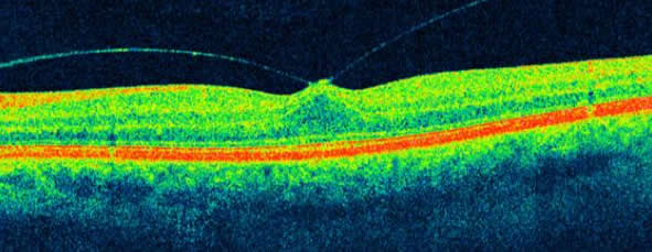
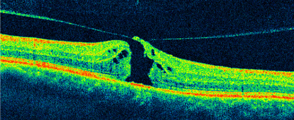
As this continues, there is more advanced forward movement of the central macular or foveal tissue.
The retina is multi-layered and ongoing traction (“pull”) from the jelly can result in a swelling of the macular area as the retinal layers separate. The inner layers are pulled inwards towards the vitreous cavity whilst the outer layers remain adherent to the eye wall. The situation worsens as normal eye movements cause the jelly within the eye to rotate, this movement producing a relentless pull on the macula. Treatment of this condition involves removing the vitreous jelly. This is called a vitrectomy. The separation of the vitreous from the macula allows the layers to return to a normal position. The damage caused by swelling in the macular can be permanent if left untouched so early surgery is often advisable.
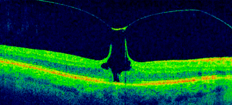
If this forward movement of the retinal tissue continues as in the following 2 pictures, the vitreomacular traction can progress to a full thickness macular hole. In this case all the tissue including the photoreceptors have been displaced, resulting in a central blind spot acutely appearing in the patients vision.. Obviously, it is always better to intervene before it gets to this stage.
If the macular hole does occur, then macular hole surgery is possible in many cases. The displaced tissue is replaced in the damaged area, with varying degrees of visual improvement. After macular hole surgery the vision is almost always below normal. This is why early intervention of agressive Vitreo Macula Traction is preferable in most cases. This picture shows the retinal appearance after successful macula hole surgery.
The common symptoms of vitreomacular traction are listed below
- appearance of a blurred patch in the centre of vision.
- distortion of straight lines to appear bent or broken
- greying of central vision
- loss of colour vision
Often these symptoms are more obvious when the patient is focussing on close tasks such as reading a book. The reason for this is that the crysalline lens moves forward during close focussing, encouraging the vitreous jelly to do likewise, leading to more vitreo macula traction.
An Amsler chart is a good way to detect early distortion in your central vision, or a change in size of the area of blurred or greyed vision.
These symptoms are not unique to Vitreo Macula Traction, but can occur in other conditions affecting the central retina such as Dry Macular Degeneration,Wet Macular Degeneration and Macular Dystrophies.
A sudden change in your central vision requires an urgent eye exam by an experienced optometrist or ophthalmologist and OCT scanning. OCT scanning is available at all Matheson Optometrists practices.
Locally we have an vitreo retinal specialist surgeon called Andrew Luff. He practices at The Optegra Eye Hospitals in Fareham and Guildford and the Wessex Nuffield in Chandlers Ford.